CASE REPORT | https://doi.org/10.5005/jp-journals-10005-1729 |
Multidisciplinary Management of Chronic Atypical Facial Pain of Psychogenic Origin: A Unique Case Report
1–5Department of Paedodontics and Preventive Dentistry, Vydehi Institute of Dental Sciences and Research Centre, Bengaluru, Karnataka, India
Corresponding Author: Jaya Naidu, Department of Paedodontics and Preventive Dentistry, Vydehi Institute of Dental Sciences and Research Centre, Bengaluru, Karnataka, India, Phone: +91 9845402319, e-mail: jayanaidu@hotmail.com
How to cite this article Naidu J, Bhattacharya P, Mendonsa JP, et al. Multidisciplinary Management of Chronic Atypical Facial Pain of Psychogenic Origin: A Unique Case Report. Int J Clin Pediatr Dent 2020;13(2):196–198.
Source of support: Nil
Conflict of interest: None
ABSTRACT
Aim:: The following case report discusses the diagnostic dilemma presented by and the multidisciplinary management of a patient with chronic atypical facial pain of psychogenic origin.
Background:: Though oral health care professionals are primarily concerned with the treatment of somatic disorders of the orofacial region, there remains a particularly challenging need to identify, diagnose and treat various psychological and psychosomatic symptoms.
Case description:: This case report describes the management of a 13-year-old male patient with the chief complaint of pain and discoloration of the skin over the right side of the face for the last 5 months, who also demonstrated symptoms of chronic anxiety and social withdrawal. As no associated soft or hard tissue abnormalities could be identified, a diagnosis of atypical/psychogenic facial pain was established. The skin discoloration was diagnosed as pityriasis versicolor and treatment for the same commenced. Following the complete resolution of the skin lesion the patient was asymptomatic, and no longer anxious.
Conclusion:: Our role as dentists is to mitigate the suffering of patients and to improve their quality of life in collaboration with specialists in psychosomatic medicine.
Clinical significance:: This case highlights that dentists must be trained to treat not only teeth, but also attend to patient’s psychosomatic symptoms.
Keywords: Chronic atypical facial pain, Psychogenic pain, Psychosomatic disorder.
BACKGROUND
Pain is almost always indicative of some degree of dysfunction which in most cases is physical.1 However, sometimes pain is devoid of any organic basis. Labeled as “inorganic” or “functional,” such pain results from an emotional illness and, although no peripheral tissue damage exists, is as distressing as somatic pain.2 It has been established that not all pain originates in sensory structures, and not all pain behavior reflects pain.3
Pain is a highly complex phenomenon and is often the outcome of a combination of physical and psychological causes. Pain is both a sensation and an emotion.3 Psychological factors affect the way people experience and express pain while chronic pain often results in secondary personal difficulties.2 The psychological consequences of pain often manifest as depression, anxiety, demoralization and irritability as a reaction either to the pain or to the losses of lifestyle and autonomy that result from it.
Psychogenic pain is a pain disorder associated with psychological factors. Mental or emotional problems can result in or exacerbate pain. In rare cases, psychogenic pain can stem purely from a mental ailment, though in some cases psychogenic pain occurs in response to a previous injury while in most cases, psychogenic pain causes existing pain as the result of some physical stimulus to feel more intense.4
It can be acute, manifesting only briefly, or chronic, persisting and recurring over weeks, months or years.4 Chronic psychogenic pain can result in constant discomfort in spite of medication, difficulty describing the nature of pain, worsening pain without any underlying medical condition and nonlocalized pains involving large parts of the body in the absence of any underlying chronic disorder with a physical cause.4
Many dental patients complain of oral symptoms after dental treatment, such as chronic pain or occlusal discomfort, for which the cause remains undetermined. Chronic idiopathic facial pain is a common problem with many symptom complexes of facial pain, that is, myofacial pain dysfunction syndrome, atypical facial pain, atypical odontalgia, oral dysesthesia, and idiopathic facial pain are frequently associated with other chronic pain conditions, a condition that is described as whole body pain syndrome.5
The following case report discusses the management of a patient who presented with chronic atypical facial pain of psychogenic origin.
CASE DESCRIPTION
A 13-year-old male patient was referred to the Department of Paedodontics and Preventive Dentistry with the chief complaint of pain in the right side of the face, right-sided headache, and painful jaw opening for the last 5 months.
The patient gave a history of extraction of the maxillary deciduous second molar 5 months back, reportedly without the administration of local anesthesia. The parents reported that the child was left psychologically traumatized by the experience and that following the dental extraction, began suffering from continuous pain on the right side of the face. Two months after the tooth extraction, the parents noticed a brownish patch developing on the right cheek region of the patient. The patient also gave a history of difficulty in mouth opening since 3 months and reduced oral intake of food with subsequent weight loss of 4–5 kg during the period. The patient visited several local dentists and general medical practitioners for the management of the pain and was prescribed various drugs which included analgesics, muscle relaxants, broad spectrum antibiotics, and mouth washes but showed no improvement in symptoms. The parents also reported that the child started being chronically anxious and refused to go to school and withdrew from his social circle. Patient demonstrated tense cooperative behavior on presentation (Frankl’s behavior rating: negative).
Extraoral examination revealed hyperpigmentation of the skin overlying the right cheek region. The brown pigmented lesion measured about 4 cm × 3 cm in size, with no sinus tract opening or pus discharge. (Fig. 1) There was tenderness over the right parotid and masseteric region. Examination of the TMJ revealed no abnormality. There were however no definite trigger zones. Intraoral examination of the patient revealed a permanent dentition, generalized extrinsic stains, and chronic generalized marginal gingivitis Ellis Class II fracture of 21. Tooth number 15 had erupted and no associated soft or hard tissue abnormalities were detected intraorally (Fig. 2).
The orthopantomogram revealed no discernible dental anomalies (Fig. 3). The child was referred to the Department of Radiodiagnosis and imaging for ultrasound of right cheek and Department of Neurology for neurological evaluation. As the brownish skin discoloration appeared to be superficial and dermatophytic in nature, the patient was referred to the Department of Dermatology for opinion. No sonological abnormality was detected on the ultrasound. The Department of Neurology provisionally diagnosed him to be suffering from pathology of the maxillary division of trigeminal nerve secondary to the dental extraction and prescribed Tegretol and Naproxen tablets, after which he reported some but not complete relief from the facial pain. The child also continued to be anxious and had difficulty in opening the mouth. The patient reported that despite being on pain medication, he continued to experience pain which was moderate to severe in intensity, continuous in nature, and radiated to the temporal region.
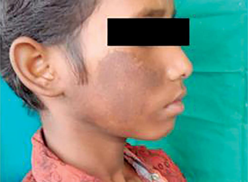
Fig. 1: Preoperative lateral view
Based on the history and clinical examination of the patient, a diagnosis of chronic atypical facial pain, psychogenic in origin, and pityriasis versicolor resulting in cutaneous hyperpigmentation was established.
The parents and the patient were given informed reassurance, a psychological technique involving empathic explanation of the disease status, thereby alleviating a great of anxiety. During the follow-up appointments, the patient received positive reinforcement and was managed with tender loving care (TLC) approach. Oral prophylaxis and L.C composite build-up for 21 was done. For the skin lesion, the patient was prescribed Ketoconazole Soap (2%) following the use of which there was complete resolution of the brownish pigmentation of the skin of the right cheek within a few days. With the resolution of the skin discoloration, the patient reported significant improvement in symptoms despite not taking any pain medication (Figs 4 and 5).
REVIEW AND FOLLOW-UP
On final review, 7 days after completion of treatment, the patient was asymptomatic and was no longer being anxious and presented a cheerful demeanor and cooperative behavior (Frankl’s behavior rating: positive).
DISCUSSION
Early identification and diagnosis of psychogenic pain ensures that ineffective and needless treatment is not administered to the patient as the same can worsen the patient’s mental state and prove to be extremely discouraging to the physician.2 Patients with AFP often consult numerous dentists and physicians seeking an explanation and effective treatment. Their use of medical and dental services is excessive, costly and usually unsatisfactory. A history of multiple ineffective treatments is common. Behavioral and psychological abnormalities are often present in AFP but are likely to be a consequence of chronic pain. Behavioral and psychological abnormalities are part of chronic pain disorders regardless of the original source or site of pain.6
In chronic pain, the principal tool for diagnosing pain is the patient’s behavior, which includes verbal communication. Other diagnostic clues include developing familiarity with known conditions, consistency of complaints and dysfunction over time and situation, consistency of pain behavior with anatomy and physiology, and collateral information from other evaluating professionals and relatives.3 Long-standing pain without specific aggravating or relieving factors, and without any radiological or neurological signs, is classified as pain of psychogenic origin that is lacking any organic structural origin.4
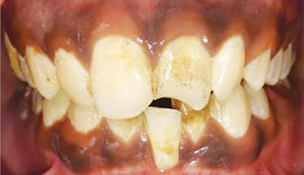
Fig. 2: Preoperative intraoral view

Fig. 3: Orthopantomogram

Fig. 4: Postoperative lateral view

Fig. 5: Postoperative intraoral view
The International Association for the Study of Pain recommend that if a patient experiences pain for psychological reasons and if they report it in the same ways as pain caused by tissue damage, it should be accepted as pain.7
The diagnosis of psychogenic pain should also never be used as an excuse to disregard the patient’s symptoms or be dismissive of the patient’s suffering. Once a diagnosis has been made, patients should be managed with a multidisciplinary approach with pain specialists, therapists, or psychiatrists and any other required specialists collaborating closely with the patient to treat both the physical and mental causes of their conditions.4
An effective treatment plan with the objectives of reduction in pain, increase in the ability to function, improved coping skills, reduction in stress and return to normal activities should be initiated.
In the present case due to the pain and psychological trauma experienced by the child during the extraction, the child developed chronic facial pain. The concurrent development of a cutaneous fungal infection resulting in hyperpigmentation of the skin resulted in the child’s dysphoria. But clinically there was no correlation between the complaints and a clear-cut association with psychological stressors. A multidisciplinary approach was adopted leading to the complete resolution of the patient’s symptoms. Referral to the Department of Psychiatry for counseling was under consideration; however, the same was deferred on account of patient being symptom free.
CONCLUSION
Though psychogenic pain is experienced as real as that produced by the known pain transmission pathways, the concept of psychogenic pain has always been controversial. Faced with the challenge of demonstrating the reality of an invisible condition, psychogenic pain is often misdiagnosed and poorly managed. The greatest challenge is however to validate the patient’s account of his/her disease without necessarily accepting their understanding of their condition.
CLINICAL SIGNIFICANCE
Most patients with psychogenic facial pain disorder report first to dentists who may then be required to identify and manage the pain while making a referral to a mental health professional or specialists in psychosomatic medicine. We also need to reassure the patient that no stigma is attached to seeking psychological help. Our role as dentists is to mitigate the suffering of patients and to improve their quality of life in collaboration with specialists in psychosomatic medicine. Thus, the need to educate dentists to address not only oral health concerns but also the patient’s psychosomatic discomfort has become an important priority.
REFERENCES
1. Wyke B. The neurology of low back pain. In: Jayson MIV. The Lumbar Spine and Back Pain,Kent: Pitman Medical; 1980. p. 265.
2. Ombregt L. A System of Orthopaedic Medicine, Elsevier; 2013.
3. Covington EC. Psychogenic pain—what it means, why it does not exist, and how to diagnose it. Pain Med 2000;1(4):287–294. DOI: 10.1046/j.1526-4637.2000.00049.x.
4. Chhabria A. Psychogenic pain disorder-differential diagnosis and treatment. J Assoc Physicians India 2015;63(2):36–40.
5. Toyofuku A. Psychosomatic problems in dentistry. Biopsychosoc Med 2016;10(14):1–5. DOI: 10.1186/s13030-016-0068-2.
6. Bhatia NK, Bhatia MS, Grewal H, et al. Atypical facial pain and depression. Delhi Psychiatr J 2012;15(1):219–220.
7. Treede RD. The international association for the study of pain definition of pain: as valid in 2018 as in 1979, but in need of regularly updated footnotes. Pain Rep 2018;3(2):e643. DOI: 10.1097/PR9.0000000000000643.
________________________
© The Author(s). 2020 Open Access This article is distributed under the terms of the Creative Commons Attribution 4.0 International License (https://creativecommons.org/licenses/by-nc/4.0/), which permits unrestricted use, distribution, and non-commercial reproduction in any medium, provided you give appropriate credit to the original author(s) and the source, provide a link to the Creative Commons license, and indicate if changes were made. The Creative Commons Public Domain Dedication waiver (http://creativecommons.org/publicdomain/zero/1.0/) applies to the data made available in this article, unless otherwise stated.